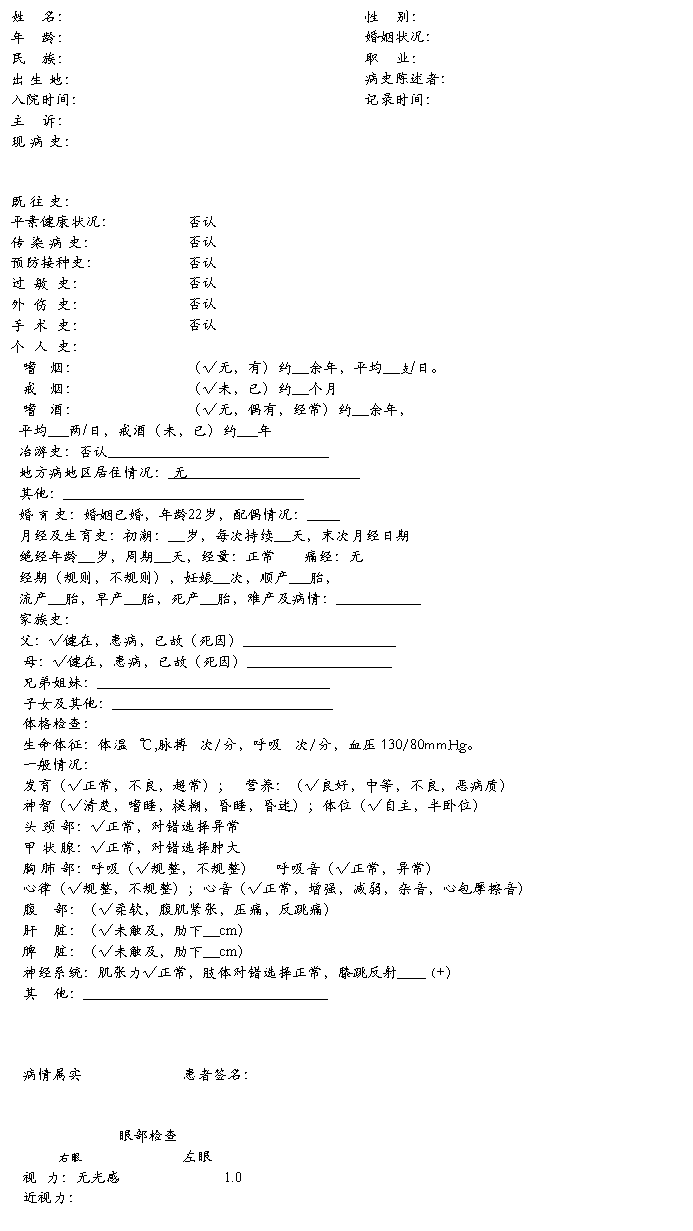
眼科病例模板

眼 位:√正常,内斜,外斜 度 √正常,内斜,外斜 度
眼球运动:√正常,转动受限 √正常,转动受限
眼 睑:√正常水肿斑痕内外翻 √正常水肿斑痕内外翻
睫 毛:排列(√整不整),倒睫 排列(√整不整),倒睫
睑结膜:√充血乳头滤泡异物 √无充血乳头滤泡异物
球结膜:正常 √充血,颞上见睑球粘连 √正常 充血混合充血水肿
混合充血水肿
泪 器:压挤分泌物返流(有,√无) 压挤分泌物反流(有,√无)
泪道√通畅,阻塞 泪道√通畅,阻塞
角 膜:√正常 水肿混浊血管 √正常 水肿混浊血管
KP
巩 膜:√前节巩膜未见异常 √正常,黄染(有无)
前 房:正常 √深 浅房水混浊 √正常 深,见玻璃体 浅房水浑浊
Tyndall征 Tyndall征
瞳 孔:不圆,散大状,直径约6.0mm 正圆,药物散大,直径约6.0mm
虹 膜:纹理(√清,下方后粘连 模糊) 纹理(√清 模糊)结节
结节
新生血管 新生血管
脱色素前粘连后粘连 脱色素前粘连后粘连
晶 体:透明 √混浊伴抖动 异物 √透明 缺如 异物
脱位 脱位
玻璃体:透明 视不见 液化 √积血 √透明 浑浊 液化 积血
后脱离膜形成窥不见 后脱离膜形成窥不见


眼压:Tn 眼压:Tn
光定位:--- +++
--- +++
--- +++
红绿色觉:-红,-绿 红绿色觉:+红,-绿
辅助检查:眼部彩超(20##年12月17日)回报:右眼玻璃体积血,右眼眼底出血,增殖,牵拉,右眼眼底球壁不均匀增厚。房角镜检查回报:房角12:00-8:00后退,鼻侧睫状体离断。
初步诊断:玻璃体积血(右)
外伤性白内障(右)
晶体不全脱位(右)
房角后退(右)
睫状体离断(右)
第二篇:英文眼科病例模板描述
1、A Discribe Sample
A vertical OCT tomogram is acquired through the macula which shows from above down neurosensory retinal detachment to the inferior fovea in the scan fields. The fovea is obviously elevated to 1000μm,
2、ACUTE RETINAL NECROSIS
OCT shows cystoid macular edema and diffuse outer retinal edema and exudates.
3、AION
The papillary swells and is elevated obviously. The physiologic cup slmost disappears. The peripapillary retinal nerve fiber swells, the thickness of which is increased obviously.
4、BECHET
There is cystoid edema in the macula, with well-defined detachment of neurosensory retina in the fovea and diffuse periphery retinal edema. (serous detachment of neurosensory retina ) Retina in the fovea is thinned. Sporadic hyperreflective points due to the exudates of hard lipids shadows the reflection from the tissues below. The margin of the optic disc is elevated obviously, which represents papilledema. ( papilledema) The retina in the
fovea is thickened. Hyperreflective band just anterior to the neur
5、BEST DISEASE
RPE/choriocapillaris in the macula is elevated, in deeper layer of which there is moderate reflective band.(RPE solid elevation)There is serous pigment endothelium detachment in the superior of vitell
6、CENTRAL SEROUS CHORIORETINOPATHY
There is serous retinal detachment in the macula, with retinal edema or cystoid edema. The neurosensory retina is elevated in the fovea, with the thickness of . Liquid dark area exists below. The RPE/choroicapillaris reflective band is clearly visible or damaged. The RPE may be elevated and detached.
7、CHOROIDAL HEMANGIOMA
Retina is sphere-like elevated. Neurosensory retinal detachment is visible in the margin of the tumor. The reflective band of RPE/choroidocapillay is disordered,only sporadic and thin choroidal reflective bands are visible in below. The retina is elevated, with serous retinal detachment around the tumor, intraretinal fluid in the retina above the tumor. (RPE damage) There is
intraretinal department with tissues connecting in between in the retina above the tumor. There is shallow retinal detachment in the
8、CHOROIDAL OSTEOMA
The RPE/choroid reflective band is enhanced and broadened irregularly, partially elevated and breaks in temperal papillary. The shallow neurosensory retinal detachment is observed in the macula, (neurosensory retinal detachment). The retina in the fovea is thinned, The retina is intraretinal departed with reflection of tissues in between, representing secondary retinoschisis in the macula and papillary, with disordered reflection.
9、CNV
The strongest reflective band (RPE/CC) ruptures. There is a multilayer hyperreflction subretinal elevation in the rupture. Neurosensory retinal detachment, retinal edema and exudation are visible.
10、CONTUSION OF RETINA
There is full-thickness retina loss in the macula, with surrounding neurosensory retina edema. (macular hole and edema) Moderate reflection in the fovea and the both sides hyperreflection of hemorrhage are observed when
RPE hemorrhagic detachment exists. The choroidal reflective band is enhanced in the temporal fovea, which represents choroid rupture.(choroid rupture with hemorrhagic RPE detachment) The RPE/choriocapillaris reflective band is broken and disordered in the temporal fovea, with the reflective
11、DIABETIC RETINOPATHY
(macular edema)The neurosensory retina shows thickened thickness and diffuse reduced interlaminar reflection in the macula and periphery retina. There are sporadic hyperreflective points in outer retina shadowing the reflection returning from below, due to hard exudates. The serous neurosensory retina detachment exists in the macula, with detachment cavity shown as fluid dark area. The local elevation of retinal nerve fiber layer shows enhanced reflection and shadows the reflection returning from below, wh
12、Drusen
Hard drusen shows the local elevation of RPE and tissues below, with hyperreflectivity. Soft drusen shows semispherical elevation of RPE.
13、Dry AMD
Hard drusen shows the local elevation of RPE and tissues below, with hyperreflectivity. Soft drusen shows semispherical elevation of RPE.
RPE/choriocapillaris is elevated well-defined. (hard drusen) The neurosensory retina is normal or thinned in corresponding area. The reflection band of RPE/choriocapillaris is elevated like semisphere or merged-semisphere. There is moderate density reflection band below, connecting with the choroidal reflection band. (soft drusen) . Retina above is thinned, while the reflection band of RPE/choriocapillaris is enhanced.(choroidoretinal geographic atrophy). The RPE reflection band disappears somewhere.(RPE a
14、EPIRETINAL HEMORRHAGE
Retina seems to be elevated, with dense hyperreflection anterior to the retina. All the reflection from tissues behind it disappears. It’s hard to identify the hemorrhage is located under the inner limiting membrane or behind the posterior limiting membrane of the vitreous.
15、EPIRETINAL MEMBRANE IN THE MACULA The epiretinal membrane appears as a streaky-like enhanced reflective band just attached anterior to the
neurosensory retina. The depression of the fovea disappears, and macular edema forms. The thickness of the retina is increased in the fovea, with steep contour and pseudohole forms.
16、EPIRETINAL MEMBRANE
The moderate and high reflective band is shown adhered tightly anterior to the retina, which may track the retina, resulting in retinal pucker and retinal edema.
17、GLAUCOMA RETINAL NERVE FIBER LAYER The reflection of retinal nerve fiber layer is thinned diffusely.
18、GLAUCOMA
The physiologic cup is enlarged and deepened, the reflective band is thinned or breaks in the superior cup wall. The cup is enlarged.
19、HARD EXUDATION
Potted and sheet hyperreflection is shown in outer plexiform layer, attenuating the reflection behind.
20、IDIOPATHIC CNV
Retina swells and is thickened. Serous retinal detachment is shown, while choroidal neovascular is visible under the macula.
21、IDIOPATHIC CNV-1
There is cystoid edema in the fovea. The hyperrefletive points in the outer retina which attenuate the tissues behind represent the hard exudation. Hemorrhagic or serous neurosensory retinal detachment forms. The RPE/choriocapillaris reflective band is fusiform-like enhanced in the upper macula, consistent with choroidal neovascular. The topography map shows that the retina is thickened, white and red, in the fovea and above (correspongding the CNV) .
22、JUVENILE RETINOSCHISIS
Cystoid alteration is seen in the macula, and the cavity is departed by tilted and vertical tissues. Peripheral neurosensory retina shows intraretinal department, with column tissues connecting in between. The thickness of the retina is increased in the fovea, especially in the inferonasal retina, with the superotemporal retina thinned. (cystoid macular edema). The inner wall ruptures after cystoid alteration in the macula, then the lamellar hole forms.(lamellar macula hole) . Neurosensory retinal detachme
23、MACULAR HOLE
Full-thickness hole: loss of full-thickness retina shows no reflection. Lamellar hole: The loss of the inner retina, part of the reflection is absent. stage I macular hole shows disappearance of the normal foveal contour and a low reflection field in below , but the inner layer of the retinal doesn’t break in the macula. Vitreous traction to the fovea is visible. Alleviation happens spontaneously in some cases. Stage II shows the breaks of inner surface and small full-thickness loss of the retina, accounti
24、MELANOMA OF CHOROID
The retina shows a flat and uneven elevation, the reflective band of the RPE/choriocapillaris is enhanced mildly, while the reflective band of the retina is almost normal. The pigment eddothelium is elevated in the fovea, with serous retinal detachment and disorders of the reflective band of RPE above the fovea. (RPE damage) Neurosensory retina is elevated and detached. (retinal detachment).
25、NORMAL MACULA
The thickness of the retina of the fovea is mm. There is no significant abnormality in the macular contour.
26、NORMAL OPTIC DISC
The superior, inferior and nasal margin of the optic disc is elevated mildly. The physiologic cup is small and shallow. The depression and the slope are symmetric. The moderate reflective band representing the tissues before the cribriform form is visible in the bottom of papillary, under which is the hyperreflective band of the cribriform plate. (normal optic disc).
27、OPTIC DISC PIT
The vertical scan through the optic disc shows a dark area without reflection, because of loss of cribriform plate in the inferior papillary, which represents optic disc pits. Horizontal scan shows loss of cribriform plate in the temporal optic disc, which connects with outer retinal retinoschisis and edema in the macula. Neurosensory retinal detachment exists in the macula, and there is only inner tissue with thin-wall, which represents outer wall hole. (optic disc hole and retinal edema and retinoschisis
28、OPTIC NERVE ATROPHY
The normal elevation of the papillary margin disappears. The physiologic cup shallows, with thinning of the peripapillary retina and reflection band of the retinal nerve fiber layer.
29、OPTIC NEURITIS
(epiretinal hemorrhage) The reflection in front of the retina is enhanced and attenuates, while hemorrhage is shaped in fluid level, attenuating the tissues below. The inner limiting membrane departs from the retina in superior of hemorrhage level.(intraretinal hemorrhage)The retina is elevated where retina hemorrhage exists. The intraretinal hemorrhage is shown as dark area, attenuating the reflection from below. The retinal detachment exists above the hemorrhage field.(subretinal hemorrhage) neurosensory
30、Papilledema
The margin of the papillary is moderately or mildly elevated. The physiologic cup gets shallow and elevated. (mild edema) Papillary is elevated obviously like mountain, and the cup almost disappears. (obvious edema) The papillary is elevated highly, and the cup disappears. (high cranial pression papilledema) The papillary is elevated, with papilledema, peripheral retina edema, cystoid macular edema, obvious retina edema in the posterior pole.(stasis retina edema) The papillary gets edema, with margin eleva
31、PATHOLOGIC MYOPIA
OCT shows thinning of the neurosensory retina in the central macula. The intralamellar department exists in the outer retina and inner retina, with columns connecting in between. The reflectivity of outer neurosensory retina in the inner side of RPE is visible (secondary retinoschisis) , as well as detachment of neurosensory retina. Neurosensory retina in the macula losses partially and epiretinal membrane forms.(retinoschisis and lamellar hole)OCT shows loss of full-thickness neurosensory retina in the m
32、PCV
Polypoidal choroidal vasculopathy shows cone-like steep elevation. There is thin discontinuous streaky-like hyperreflective band departing from RPE in the side of elevation, which is consistent with polypoidal choroidal vasculopathy. The neurosensory retina is thin above the elevation. Moderate reflective points are visible below the elevation, attenuating the reflection from choroid. There is neurosensory retinal detachment, with exudation in outer retina and hemorrhage pigment endothelium detachment besi
33、PIGMENT ENDOTHELIUM DETACHMENT
Retinal pigment endothelium is elevated, with fluid dark area or reflective points below, shadowing the reflection from bruch membrane and choroid.
34、POSTERIOR VITREOUS DETACHMENT
A moderate reflective band departs with the retina, floating in the posterior vitreous cavity like an irregular curve or a semiarc.
35、RAO
(In the early stages of CRVO) There is cystoid macular edema, uncomplete posterior vitreous detachment tracking the macula. Retina shows edema and enhanced reflection, is thickened. The dark area of photoreceptor is broadened. (CRAO edema recedes)The cystoid edema in the macula recedes, uncomplete posterior vitreous detachment is thinned and weak. The dark area of photoreceptor is broadened. (CRAO atrophy stage)the thickness of the retina in the centre is mildly thinned, uncomplete posterior vitreous detac
36、REDIATION RETINOPATHY
There are retinal cystoid edema ,intraretinal exudation and neurosensory retinal detachment in the macular,
(neurosensory retinal detachment)OCT shows serous neurosensory retinal detachment, neurosensory retinal cystoid edema and diffuse edema in the macular.(papilledema)papillary elevates, the physiologic cup almost disappears.(retinal exudates)Well-defined edema and elevation of retinal nerve fiber layer consistent with the soft exudates attenuates the reflection from below. The exudates appear as the
37、RETINAL ANEURYSM
(epiretinal hemorrhage) The reflection in front of the retina is enhanced and attenuates, while hemorrhage is shaped in fluid level, attenuating the tissues below. The inner limiting membrane departs from the retina in superior of hemorrhage level.(intraretinal hemorrhage)The retina is elevated where retina hemorrhage exists. The intraretinal hemorrhage is shown as dark area, attenuating the reflection from below. The retinal detachment exists above the hemorrhage field.(subretinal hemorrhage) neurosensory
38、RETINAL DETACHMENT
Neurosensory retina is elevated. The fluid below shows liquidity dark area with non-reflection, or potted or sheet
moderate or high reflection in the liquidity dark area.
39、RETINAL DETACHMENT-1
Rhegmatogenous retinal detachment (mild and moderate retinal detachment in the macula)Retinal detachment exists in the macula and inferior retina, with retinal edema. (highly retinal detachment in the macula)highly retinal detachment exists in the macula, OCT cann’t show RPE/choriocapillaris reflective band.(neurosensory retinal department)Outer and inner neurosensory retinal departs。 (wavy alteration of retina)Outer neurosensory retina shows wavy alteration (neurosensory retinal edema)Outer retinal shows
40、RETINAL EDEMA
Retina shows cystoid alteration in the fovea, with reflection in the cavity reduced and inner surface of the retina elevated.(with the height of mm) The retina is thickened diffusely in the macula. The reflection of the outer retina is influenced, and the IS/OS and RPE reflection is decreased, because of the retinal edema.
41、RETINITIS PIGMENTOSA
The macula shows cystoid edema, with large sac, retinal pigment endothelium proliferation and migration towards
the inner retina. The retina is thinned, while the RPE/choriocapillaris reflective band is sporadicly enhanced. (migration and proliferation of RPE).
42、RETINOSCHISIS
The reflection of the retinal focus is reduced. Outer plexiform layer is teared and enlonged, with bridge-like junction.
43、RVO
(retinal hemorrhage)【shallow retinal hemorrhage】Cystoid edema exists in the macula. The reflection of the retina is enhanced in the fovea, but doesn’t attenuate the reflection from deeper, which represents shallow retinal hemorrhage. 【deep retinal hemorrhage】The neurosensory retina superior to the papillary is elevated and enhanced, and attenuates rapidly, with dark area below and RPE/choriocapillaris reflective band shadowed.(retinal exudation)【soft exudation】There is cystoid edema in the macula and diffu
44、SOFT EXUDATION
The small occlusion focus of nerve fiber layer shows local elevated hyperflection in the retinal nerve fiber layer, attenuating the reflection behind.
45、STARGARDT
The dark area of retinal photoreceptor in the macula disappears, and the retina is thinned, the reflective band of RPE/choriocapillaris around the fovea is enhanced unevenly, the neurosensory retina in the macula almost disappears.
46、SYMPATHETIC OPHTHALMIA
There are multiple serous neurosensory retinal detachment and exudation in the macula and posterior pole.(serous neurosensory retinal detachment) The margin of the papillary is elevated mildly.(papilledema) Retina in the macula is thinned, while the reflective band of the choroid in the parafovea is enhanced and thickened unevenly.(retina thinning).
47、UVEITIS
Diffuse retinal edema exists in the fovea and temporal retina, with neurosensory retina thickened, cystoid macula edema, serous neurosensory retinal detachment and enhanced reflection of inner retina. Pigment endothelium detachment exists, with spongy retina edema. The margin of the optic disc shows moderate elevation, representing papilledema. The retina is thinned
in the fovea. Outer retina shows sporadic hyperreflective points in the macula, attenuating the tissues behind, representing the hard exudatio
48、VITREOMACULAR TRACYION SYDROME The moderate and high reflective band of the vitreous posterior limiting membrane adheres with the macula, especially with the fovea. The fovea is tracked, elevated and thickened with edema.( μm) The neurosensory retina reflection from below is shadowed, and retina reflection rupture is visible sometimes due to the hole.(macular edema)
49、VKH
OCT shows multifocal serous detachment of neurosensory retina at the fovea, department of inner and outer retina in macula and papillary. Retinal detachment is shown in the macula, serous retinal detachment in the superior macular and exudative retinal detachment in the inferior macula. Moderate density exudates exist between the RPE and the detached neurosensory retina. Exudative retinal detachment is seen in the macula and inferior retina. Hyperreflective bands due to the exudates of hard lipids are seen
50、W-AMD
RPE/chorocapillaris reflective band breaks, and it is fusiform-like broadened in the subretinal fovea. The retina shows cystoid edema. RPE/chorocapillaris reflective band breaks, and it is broadened irregularly, (occult CNV) with serous neurosensory retinal detachment above. Retinal pigment endothelium is elevated with moderate and high reflection below (CNV membrane), neurosensory retina cystoid edema above. There is hyperreflective mass in the neurosensory retina, attenuating the retinal tissues behind,(
-
病例范文
检查的项目1一般项目籍贯须写明省市或县别入院日期急症或重症应注明时刻均应填年月日病情陈述者填患者如系旁人代述应说明可靠程度2主诉电…
-
住院病例范本
住院病例姓名杨树广性别男年龄73岁民族汉族出生地湖南保靖入院日期20xx0112病史叙述者患者本人可靠程度可靠婚姻已婚职业农民住址…
-
中医病历范本
医院入院记录科别康复科病床号9门诊号住院号xxxx医院科别康复科病床号9门诊号住院号xxxx医院入院记录科别康复科病床号8门诊号住…
-
内科病例范文
姓名科别住院号医保号性别工作单位年龄岁住址婚姻供史者出生地址入院日期年月日时分民族病史采集日期年月日时分主诉肝硬化10余年伴腹胀下…
-
门诊病历范本
咽异物xx年xx月xx日食鱼后咽部异物感3小时患者3小时前食鱼时出现左咽部异物扎刺感咳之不能排出异物异物感持续存在吞咽时刺痛感加剧…
-
各科病历书写范文
第一节病案书写的一般要求及注意点1新入院患者的入院记录由住院医师认真书写有实习医师者除入院记录外另由实习医师系统书写入院病历入院病…
-
内外妇儿各科病历书写范文
各科病历书写范文病案书写病案系病历及其它医疗护理文件的总称病历包括入院记录入院病历病程记录手术记录转科记录出院记录和门诊记录等第一…
-
20xx年各科病历书写范
各科病历书写范文病案书写病案系病历及其它医疗护理文件的总称病历包括入院记录入院病历病程记录手术记录转科记录出院记录和门诊记录等第一…
-
妇产科病历书写范例1
初步诊断1宫内妊娠32周妊3产0头位2妊娠高血压疾病产前子癎3慢性高血压合并产前子癎4双测视网膜脱落医生签名病程日志20xx121…
-
眼科门诊病例书写
眼科门诊记录门诊记录姓名冯**性别男年龄15门诊号910903初诊记录1991-9-3右眼红、痛、畏光、流泪3天。患儿3天前晨起感…